

 Sinem Nedime Sökücü1 , Resit Akyel2 , Fatma Tokgöz Akyıl1 , Seda Tural Önür1 , Kaan Kara1 , Nurdan Şimşek Veske1 , Fatma Elif Çayır1 , Cengiz Özdemir3
 2Deparment of Nuclear Medicine, University of Health Sciences, Yedikule Chest Disease and Thoracic Surgery Training and Research Hospital, Istanbul, Turkiye
3Liv Hospital, Vadi Istanbul

Abstract
BACKGROUND AND AIM: This study reviews the clinical characteristics, radiological findings, and diagnostic procedures for patients with pulmonary actinomycosis (PA). It also assesses the utility of 18F-fluorodeoxyglucose positron emission tomography-computed tomography (PET-CT) scans in diagnosing PA.
METHODS: Conducted retrospectively at a tertiary referral hospital, this study investigated patients diagnosed with PA between January 2012 and January 2022. Demographics, clinical and radiological findings at presentation, diagnostic steps, PET-CT findings, and the time interval to diagnosis were analyzed.
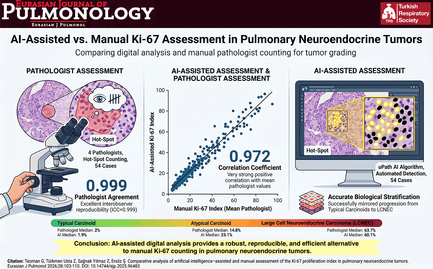
RESULTS: Among the 34 patients, the mean age at diagnosis was 49 years (range 23−77), with 19 (56%) being male. The most common symptom was cough, reported by 23 patients (68%). Chronic obstructive pulmonary disease and bronchiectasis were the most frequent underlying conditions. Typical chest tomography features included nodular lesions, mass lesions, consolidation, bronchiectasis, and atelectasis. Initial pre-diagnoses included lung cancer in 16 patients (47%), tuberculosis in 9 patients (27%), and late-resolving pneumonia in 6 patients (18%). No patient received an accurate initial diagnosis of PA. All definitive diagnoses were made histopathologically through specimens obtained from: sputum analysis in 1 (2.9%) patient, flexible bronchoscopy in 17 (50%) patients, rigid bronchoscopy in 1 (2.9%) patient, endobronchial ultrasonography in 2 (5.9%) patients, transthoracic needle aspiration in 6 (17.7%) patients, and surgical resection in 7 (20.6%) patients. The mean time from symptom onset to definitive diagnosis was 53.2±44.1 days (Range 9−175 days). Among the patients, 16 (47%) underwent PET-CT, and 10 (29%) underwent cranial magnetic resonance imaging. From the re-assessment of 13 PET-CT scans, the derived values were as follows: Standard Uptake Value (SUV) max value was 6.98±2.74 (range 0.9−9.92), SUVmean value was 3.95±1.51 (range 0.51−5.30), SUVpeak value was 5.68±2.24 (range 0.64−7.89), tumor lesion glycolysis was 138.58±151.86 (range 3−440.5), and metabolic tumor volume was 27.85±37.97 (range 0−131.00).
CONCLUSIONS: The diagnosis of PA is challenging and often delayed, frequently misdiagnosed as lung cancer or pulmonary tuberculosis. PA shows moderate metabolic uptake on PET-CT scans and, PET-CT scan is insufficient for accurate and timely differentiation between lung malignancy and PA.